


The following is information on some common conditions treated by Mr Williams. For any questions or to request a consultation please contact his office.
Patient information
Conditions treated
What is a cataract?
The lens is a clear structure inside the eye. Light is focused by the cornea (the window of the eye) through the lens onto the retina at the back of the eye. This allows us to see. Cataract is a clouding of the natural lens that can cause blurring of vision. This usually happens naturally with age but can happen sooner in life for example following injury, some types of surgery or diseases such as diabetes mellitus.
Both men and women may develop the condition and usually this starts in mid-life but gets progressively worse with age. Unfortunately, symptoms like glare and blurred vision become problematic as it may continue to progress, despite initially being managed with spectacles.
Usually, an optometrist may notice that you have cataract when they test your spectacle prescription. This sometimes results in increased myopia (short-sightedness) or a reduction in vision that glasses can no longer help. They may have told you that your vision has fallen below the legal standard for driving. Normally they will refer you for an opinion from an ophthalmologist (an eye surgeon).
The decision to consider treatment will be guided by factors such as the impact on your quality of life, job and whether you have fallen below the legal standard for driving.
What is cataract surgery?
The treatment for cataract is surgery. The aim is to remove the natural lens and replace it with an artificial lens implant. This is usually achieved by keyhole surgery through the cornea, involving the removal of the cataractous lens with an ultrasound machine called phacoemulsification. This allows us to break the lens into small pieces and vacuum these out of the eye prior to insertion of the lens implant.
When we remove the natural lens, we have an opportunity to correct distance vision. We do this by taking measurements such as the length of the eye and the curvature (steepness) of the cornea prior to surgery. This allows us to choose a lens to optimise distance vision.
What can you expect?
Surgery usually takes up to 20 minutes but may be longer in some instances. It will usually be done as a day case under local anaesthetic or eye drops. We will talk you through the steps again on the day but involves you lying flat for a few minutes once you are comfortable. Your eye will be covered with a drape while allowing plenty of fresh air.
What are the risks during surgery?
All surgery carries risk including bleeding and infection, but fortunately these are rare. We will talk you through your personalised risks prior to surgery. Approximately 1 in 10 will need some laser treatment to remove a film that may develop behind the implant (called posterior capsular opacification), usually a number of years after surgery. This can sometimes mimic the symptoms of cataract including glare and reduced vision.
Up to 1:100 will need additional surgery, for example if the lens implant cannot be inserted on the day or additional steps to remove the cataract are needed. This can include the vitreous (the jelly of the eye) being exposed, fragments of lens material becoming trapped behind the iris during surgery or bleeding within the eye. Occasionally the cornea may become cloudy during or after surgery. This may require additional surgery such as a corneal transplant if it fails to recover.
Sadly, despite scrupulous hygiene procedures and screening, there is currently an unavoidable background risk of COVID-19/SARS-CoV2 virus. Every effort is made to mitigate these risks, but we cannot guarantee that you will be free from exposure, any more than in any medical environment.
What are the risks following surgery?
Overall, most patients do extremely well, achieving good distance vision in the absence of pre-existing eye disease. You will be given drops to take home including steroid drops to help calm inflammation in the eye.
The eye will settle down in the days following surgery (see the section below on what to expect). A sensation of dryness and irritation including a foreign body sensation can be common in the first few days and usually settle down. Some patients, pre-disposed to dry eye, may have their condition exacerbated. We will usually have taken steps to mitigate this e.g. by using non-preserved drops or lubricating drops. If things are not improving it is important that we are contacted.
It is normal for the eye to take time to adjust to the change in prescription. Some patients experience glare, haloes or unusual crescentic shapes in the periphery of their vision (dysphotopsia). These usually settle down in the first few weeks, but require time, a process called neuro-adaptation. These side effects may be more common following ‘premium lenses’ (see below).
Some patients are pre-disposed to developing high pressure following surgery, more commonly when there is pre-existing glaucoma. Less than 5% of patients can also develop swelling of the macula (macula oedema) which can usually be treated with drops, but sometimes requires injections of steroid around the eye to help resolve this. It may be more common if you have pre-existing conditions affecting your macula e.g. epiretinal membranes.
Very rarely some patients, less than 1 in a 1000, may lose their sight due to a serious complication such as a retinal detachment. Although floaters may become apparent following cataract surgery, any sudden change, increase or shadows in the vision require urgent assessment to rule out a complication such as a retinal tear that can lead to a retinal detachment.
Recovery is usually fast, and most patients do not need stitches. You will usually be routinely assessed within a few weeks of surgery. Sometimes you will need to be monitored more closely.
It is imperative that should you develop any problems including reduced vision, pain or increased redness you seek help urgently. This may indicate infection (endophthalmitis). While extremely rare (again, less than 1 in a 1000) it is very serious and requires urgent/same day assessment and treatment.
Will I need glasses afterwards?
The aim of cataract surgery is to remove the cataractous lens and replace it with a clear, artificial lens implant to correct for distance in most instances. Occasionally patients may prefer to be left short sighted, for example if we are only planning surgery on one eye, to make sure the vision is balanced.
In all instances, although we are trying to reduce spectacle dependence, we cannot guarantee that you will not need glasses for support. You may need glasses to help with some distance activities if a ‘conventional’ monofocal lens for distance is chosen and may also be the case with ‘premium lenses’ (see below). This is because there is a small margin of error. 90% of patients will be within 1 dioptre (D) of the target and 70% within 0.5D. Standard lens implants, however, do not correct astigmatism (see below).
You will need help in the form of reading glasses if correcting you for distance.
What if I have astigmatism?
Astigmatism is a condition when the natural steepness of the cornea (the window of the eye) is greater in one meridian (plane) than the other. This means that a conventional lens will not be able to fully correct the way light is distorted when it enters the eye. Usually, your optometrist will ensure your glasses have a cylinder incorporated into them to account for this or contact lenses may similarly have a ‘toric’ component.
Pre-existing astigmatism will not be resolved by cataract surgery alone. This would mean that you might still need glasses or in some circumstances contact lenses to correct distance vision after surgery, even when the lens is now clear again. We will talk to you about additional steps that may be appropriate to try and reduce or overcome astigmatism during surgery such as corneal relaxing incisions or specialist lenses such as toric lens implants.
Toric lens implants are calculated by the same measurements we take for conventional cataract surgery to take account of the degree and direction of your astigmatism. This allows us to minimise the need for distance glasses, similar to a way a cylinder would be incorporated into spectacles. We will carefully screen you to see whether the type of astigmatism you have is suitable (regular), your pupils are of an appropriate size and exclude other conditions such as dry eye and other pre-existing conditions.
Toric lens implants at the time of cataract surgery have to be placed in a precise orientation in order to achieve their effect. This may mean marking the eye and/or using imaging technology to determine this orientation. The stability of these lens, that is their ability to maintain their correct orientation, is usually high with the majority rotating less than 5 degrees. This is usually well tolerated and would not generally require any intervention to re-orientate the lens, but there remains a small risk that the lens will need adjusting. Furthermore, there may be some residual astigmatism (usually less than 1 Diopter) meaning that some patients would still need spectacles to maximise their vision.
Is it possible to have a range of vision after cataract surgery?
As the natural lens becomes older, its ability to focus light on close up objects diminishes. This is what we call presbyopia and is the reason we need reading support as we approach middle age. There are a number of factors that prevent us from restoring vision to the level we enjoyed as children or young adults, and the use of ‘plastic’ lens implants is one such limitation as they are unable to accommodate i.e. change shape to bring objects close up into focus, such as when we read. There are approaches however that can help bypass some of these limitations.
Monovision
Although a conventional monofocal lens can help for distance, one will still need spectacles to help with close up activities. Some choose to wear progressive or multifocal lenses after cataract surgery following this type of lens implantation. Some patients may benefit however from a procedure called monovision. This is where we would aim to leave the non-dominant eye a little short sighted (myopic) in order to help with reading activities. Not everyone is suitable for this approach and would need to be discussed at consultation.
Other patients may benefit from lenses that enhance the depth of focus. These multifocal lenses, sometimes called ‘premium lenses’ may be an option. You will be assessed for your range of needs, your suitability including modern imagining. We will speak to you about the usual risks of surgery and the additional risks of glare and haloes that may occur with these lenses. The alternative approach to providing a greater range of vision is the use of extended depth of focus (EDOF).
EDOF lenses
EDOF lenses provide an enhanced depth of vision, helping with middle range activities e.g. using a tablet, in addition to allowing good distance vision. Although they will not usually provide spectacle independence for reading, they can be helpful for some patients who want more flexibility and may be combined with ‘mini-monovision’ to further assist with reading. They do not however eliminate the need for spectacle help, in particular for prolonged reading and/or small print.
Multifocal lenses
Multifocal lens implants (usually tri-focal), similar to incorporation in spectacles of ‘progressive’ or ‘varifocal’ lenses, split the light entering the eye into two or more images. Usually this helps with intermediate vision tasks as in EDOF lenses and also for close up activities such as reading. Some patients find this gives them a greater range of vision and reduce spectacle dependence, typically in up to 80-90% of patients. The trade off with this type of lens is obviously some degradation of the quality of vision as the image is split on entering the eye.
Both EDOF and multi-focal lens implants may need to be combined with a toric lens to reduce astigmatism, to achieve the best outcomes. This is something that would be discussed at your consultation.
Risks of premium lenses
The usual risks of cataract surgery apply to premium lenses including toric, EDOF and multifocal implants. If surgical complications occur, it may not be possible to place a premium lens as intended, and a monofocal lens may have to be used instead.
Specific risks include glare and haloes around point light sources are present in most patients, especially at night. With time, a process called neural adaptation occurs to overcome these effects and this reduces to a level that is not troublesome for the majority of patients. It can unmask itself in the evenings or in dim light conditions but may be severe for up to 1 in 6 patients. We will screen for risk factors that increase this possibility but cannot guarantee it will not occur. Rarely less than 1 in 100 may elect to have their lens exchanged (which is usually more challenging that conventional cataract surgery) or need laser surgery to modify the outcome (less than 1 in 20).
Multifocal lenses are more likely to work if used in both eyes and it is important to understand that the process of neuroadaptation is more challenging in over 70s. The loss of distance contrast, floaters, retinal detachment, dry eye and posterior capsular opacification is also higher with these lenses.
Special Circumstances in cataract surgery
If you have a very high near or far sight correction (refractive error), prior to surgery and you have cataracts in both eyes, then the conventional approach of correcting distance vision will lead to a period of imbalance before the second eye surgery takes place. We may prefer to undertake surgery on separate days but will try to undertake the second eye operation as soon as possible. Sometimes if you have a high refractive error and cataract in one eye only, following discussion, you may choose to remain far or near sighted to prevent problems with imbalance.
Previous laser refractive surgery presents a different but increasing challenge as conventional measurements taken to choose a lens implant are less predictable. We will usually talk to you about getting hold of information relating to your previous surgery including spectacle correction, the type of laser surgery and the outcome. This will give us the best chance of getting you to see glasses free afterwards. It is often helpful if you can get hold of this information prior to referral. It is important to emphasise that the outcomes are less predictable following previous laser refractive surgery.
Some patients have pre-existing corneal diseases, such as keratoconus or corneal transplantation, that makes lens selection and overcoming astigmatism more challenging. We will talk to you about this in more depth prior to surgery.
Some patients have a pre-existing vulnerability in their cornea (the window of the eye) such as Fuchs’ Endothelial Corneal Dystrophy. In this condition the endothelium (the back layer of the cornea) is not working properly and the liquid within cannot be pumped out of the cornea adequately and it becomes waterlogged. We can often detect this vulnerability even if the cornea is working reasonably well beforehand. The use of phacoemulsification ultrasound during cataract surgery may cause damage to the cells of the endothelium and result in decompensation of the cornea following cataract surgery, even if uneventful. We will talk to you about the risks and approaches including cataract surgery alone and consideration for the possibility of planned corneal transplantation (endothelial keratoplasty) if we feel the cornea is unlikely to remain clear following cataract surgery.
Anaesthesia
Broadly there are three approaches we would consider in order to numb your eye and undertake cataract surgery:
- Topical anaesthetic drops with/without the injection of an anaesthetic into the front chamber of the eye during the procedure. You will be awake for the procedure. This has the advantage of being faster and allows the eye to recover more quickly after surgery. It is important to note that your eye can move with this approach and we will encourage you to look at the light during surgery. This approach can rarely cause problems with the cornea or have unwanted systemic effect such as lowering your heart rate. This is exceptionally rare.
- Sub-tenon anaesthesia. You will be awake for the procedure. This involves the creation of a small pocket in the membrane covering the eye (conjunctiva) and the injection of an anaesthetic agent around the side of the eye to numb it. This has the advantage of lasting longer which can be helpful in more challenging cases. The disadvantages are that it may cause swelling or bleeding around the eye and can, very rarely, cause a more widespread effect including putting you to sleep. We would normally have an anaesthetic-trained colleague to undertake or be on standby for this exceptionally rare eventuality.
- General anaesthesia. Very rarely there may be medical reasons why it is advisable to be put to sleep for the procedure or you may feel unable to be awake. The risks and benefits of this approach would need to be discussed with an anaesthetist beforehand and subject to underlying medical conditions.
We may also offer you sedation to help relax you during surgery. This will depend on your medical background and personal needs and would involve taking a tablet beforehand.
Information for after your operation
After you have had an operation to remove a cataract, it is likely that your vision will be blurry until the following day due to the fact that you have just had surgery and also due to your pupil being dilated. Patients who have had cataract surgery usually comment how bright the world is even shortly after the operation.
Mild discomfort is normal, which should respond well to paracetamol alone. We would normally place a shield and ask you to keep this on overnight for the first 2-3 days or at night for a few days.
Please make sure you use the drops supplied, as directed for a total of 4 weeks, unless otherwise advised. It is often easier for someone else to put the drops in. Please ensure that hands are washed before using the drops and try not to touch the end of the bottle on the eye or surrounding skin.
Other eye drops. If you use e.g. glaucoma drops, then it is usually safe to continue, although it is important to use a new bottle for the freshly operated eye. We will let you know if we want you to stop taking any regular drops.
Showering / washing. To reduce the risk of infection, keep the eye closed in the shower and avoid splashing water into it for at least a week. We do not advise swimming for at least a month after surgery.
Exercise. You have just had an operation, so please keep exercise light for a week or so. Gentle gardening is fine, provided that at no point you bring a dirty hand near to your face / eye, as is walking the dog / golf etc.
Do not be concerned if the eye is red during the first few days, especially under the upper lid – this is normal and will settle down.
Cataract surgery changes your spectacle prescription: to allow reading in the short term, off-the-shelf ‘+2.5’ reading glasses may be worth a small investment.
Your vision should continue to improve from the first day. Should it deteriorate, it is important that you ring and let us know without delay. We will provide contact details, including out of hours numbers.
Costs
Full costs of surgery will be discussed beforehand. Typically for self-funding patients this will include the cost of an anaesthetic and your first follow up visit. The cost of a monofocal lens is lower than for toric, extended depth of focus, multifocal and multifocal toric ‘premium’ lens implants. Insurers will not typically fund these premium lenses, and if you are a suitable candidate the cost of top-up fees will be discussed with you.
The cornea is a window of transparent tissue at the front of the eyeball. It allows light to pass into the eye and provides focus so that images can be seen. Various diseases or injury can make the cornea either cloudy or change shape. This prevents the normal passage of light and affects vision.
Why do I need a corneal transplant?
Wearing glasses or using contact lenses may overcome many diseases affecting the cornea. Sometimes these approaches do not help or patients may not be able to tolerate contact lenses. In these instances it may be necessary to replace part or all of your cornea through surgery by undertaking a corneal transplant called a graft or keratoplasty. This graft will come from a donor who has died and given consent for their cornea to be used for transplantation after their death.
What types of corneal transplant are there?
Different diseases may affect all or part of the cornea. There are three broad approaches
- Penetrating Keratoplasty (PK) – a full thickness corneal transplant (Figure 1)
- Deep Anterior Lamellar Keratoplasty (DALK) – a partial thickness corneal transplant replacing the epithelium and front part of the stroma up to 90% depth (Figure 2)
- Endothelial Keratoplasty e.g. Descemet Stripping Automated Endothelial Keratoplasty (DSAEK) or Descemet Membrane Endothelial Keratoplasty (DMEK) – a partial thickness corneal transplant replacing the endothelium (Figure 3)
All involve the replacement of some or all of your cornea. Like all organ transplants, this involves taking corneal tissue from someone who has died and donated their organs for transplantation.
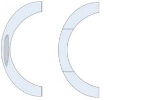
Figure 1: Penetrating Keratoplasty. A side profile cartoon showing a scar involving most of the cornea (Part A). Part B shows the replacement of the scar with a full thickness graft.
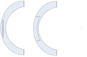
Figure 2: Deep Anterior Lamellar Keratoplasty. A side profile cartoon showing a scar involving the front part of the stroma of the cornea only (Part A). Part B shows the replacement of the front surface of the cornea with donor tissue.
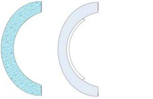
Figure 3: Endothelial Keratoplasty. A side profile cartoon showing a waterlogged cornea (Part A). Part B shows the replacement of the back surface of the cornea with donor endothelium (white).
A PK or DALK
Involves the removal of all or some of the cornea, as outlined in Figures 1 and 2 and replacement with a donor graft measuring a few millimetres. The graft must be sutured (stitched) into place and this may involve inserting several (typically 16) interrupted or continuous (usually one or two) ‘permanent’ stitches (Figure 4A or 4B). These will remain in place and will usually be completely or selectively removed several months after surgery.
These are usually undertaken under a general anaesthetic
A DSAEK or DMEK
Involves a smaller incision that allows replacement graft material to be placed inside the anterior (front) part of the eye e.g. with an insertion device which allows coiling of the graft and unfurling inside the eye, akin to getting a ‘ship in a bottle’. The graft is floated onto the back surface of the cornea with an air bubble and only a few sutures are required (Figure 4C). This procedure may be combined with cataract surgery or completed as a separate operation. For the air bubble to attach the graft you will usually be asked to lie flat on your back for an hour or so after the operation.
This is usually undertaken under a local anaesthetic.

Figure 4: Corneal suturing in different types of grafts (blue). Interrupted (Part A) or Continuous stiches (Part B) in a Penetrating/Deep Anterior Lamellar Keratoplasty. A small side incision with limited sutures is seen for an Endothelial Keratoplasty e.g. DSAEK (Part C)
What are the risks and complications of surgery?
All Grafts
All grafts carry a risk of rejection. This is because the body’s immune system may recognise the transplant as not belonging to the patient’s body. This can occur in less than 20% of patients in a PK at 2 years but is lower, around 10% for a DSAEK and lower for DMEK. Corneal graft rejection may or may not be reversible with treatment, usually steroid eye drops or tablets to dampen the immune response. All patients are given steroid drops after transplantation as a preventative measure but it is unusual for immune suppressing tablets to be given for most patients needing a corneal graft. Rejection may lead to failure of the graft i.e. it no longer works and the graft becomes cloudy resulting in blurring of vision.
Symptoms of rejection are:
- Red eye
- Sensitivity to light
- Visual loss
- Pain
If you experience any of the above symptoms do not hesitate to telephone the Spire Eye Centre during working hours or the out of hours number on 01905 350003.
Other risks include:
- Astigmatism (curvature of the eye becoming steeper in one plane after suturing)
- Glaucoma (high pressure in the eye) which can usually be managed with drops.
- Immediately after surgery: Specifically, this risk is higher in the first few days following DSAEK/DMEK due to the injection of an air or gas bubble. It is imperative that increasing pain or reduced vision is reviewed promptly.
- Long term: The use of steroid drops (necessary to prevent graft rejection) increases the risk of high pressure within the eye. This may not always be apparent to you but can lead to irreversible sight loss. This is one of the reasons we follow you up in clinic and encourage regular review at the optometrist.
- Infection. All intra-ocular surgery carries risk of infection.
- Immediately after surgery: In the days following surgery any increase in pain, redness or reduction in vision needs urgent attention. The risk of the donor material (including COVID-19 for which donors are screened) transmitting an infection is incredibly low.
- Long term: The use of contact lenses in the first few days and steroid drops to prevent rejection also increase the risk of infection, including unusual infections of the cornea. In the first few days we will provide anti-microbial drops but not usually long term. The use of steroid drops can mask the symptoms and signs of an infection and it is critical that any change in vision, pain or redness is reviewed urgently. It is also important that you inform any optometrists or doctors who may attend to any problems that you have had a corneal transplant and are using steroid drops.
- Cataract (clouding of the lens), which is amenable to surgery. However, the cataract is usually removed prior to or at the time of DSAEK/DMEK.
- Loss of sight which may be permanent, especially if unusual but serious complications such as infection, retinal detachment or bleeding occur. Sustained or high pressure may also sadly result in this complication.
- Very rarely, the eye itself may be lost if a major haemorrhage occurs during surgery
DALK
There is a risk of conversion to a full thickness graft (PK) in around 10% of cases.
DSAEK/DMEK
There is a risk of the graft becoming detached from the cornea. This happens to around 10% of patients and will usually require a further air injection to re-attach the graft.
Anaesthesia
Broadly there are two approaches we would consider in order to numb your eye and undertake corneal transplantation:
- Sub-tenon anaesthesia. You will be awake for the procedure. This involves the creation of a small pocket in the membrane covering the eye (conjunctiva) and the injection of an anaesthetic agent around the side of the eye to numb it. This has the advantage of lasting longer which can be helpful in more challenging cases. The disadvantages are that it may cause swelling or bleeding around the eye and can, very rarely, cause a more widespread effect including putting you to sleep. We would normally have an anaesthetic-trained colleague to undertake or be on standby for this exceptionally rare eventuality.
- General anaesthesia. Very rarely there may be medical reasons why it is advisable to be put to sleep for the procedure or you may feel unable to be awake for DSAEK/DMEK. We would usually consider this approach for PK/DALK due to the inherent risks of surgery. The risks and benefits of this approach would need to be discussed with an anaesthetist beforehand and subject to underlying medical conditions.
We may also offer you sedation to help relax you during surgery. This will depend on your medical background and personal needs and would involve taking a tablet beforehand.
Information for after your operation
Mr Williams will assess you following the operation and the vast majority of patients will be able to go home the same day and will be followed up in clinic within a week.
- You will be advised on specific aftercare e.g. intensity of anti-rejection drops and antibiotic drops.
- We usually advise avoiding submerging the eye, taking care to wear a shield at night or when near young children and pets and to remain off work for at least two weeks.
- We usually advise you to sleep as you would normally, with a shield on at night for the first couple of weeks. Individual circumstances can be discussed.
- After you have had an operation, it is likely that your vision will be blurry for the first few weeks. Mild discomfort is normal, which should respond well to paracetamol alone.
- A contact lens may be placed in the eye for the first few days to assist healing and comfort. We will remove this for you.
- An air bubble in the eye following surgery will dissipate in a few days. It is normal to be able to see this becoming smaller and will ‘float’ within the front part of the eye.
- You will usually be required to see Mr Williams in clinic the morning after surgery and 1 week afterwards. You will be monitored more closely in the first few weeks with outpatient appointments usually being spread out over the coming months.
- It may also be necessary to organise additional visits to an optician to help optimise your vision later on, and this may include where appropriate the use of specialist contact lenses to maximise vision. This will usually be organised following the removal of stitches, which we will usually commence a month after DSAEK/DMEK but several months later after PK/DALK.
Please make sure you use the drops supplied, unless otherwise advised. The steroid drops provided (usually Dexamethasone) are anti-rejection drops and must not be stopped or be allowed to run out unless discussed with Mr. Williams. We may consider stopping these several months after surgery.
It is often easier for someone else to put the drops in. Please ensure that hands are washed before using the drops and try not to touch the end of the bottle on the eye or surrounding skin.
Other advice
- Stop wearing your usual contact lenses prior to and after your surgery until advised to do so.
- You may feel that wearing sunglasses can make your eye(s) feel more comfortable following surgery.
- Other eye drops. If you use e.g. glaucoma drops, then it is usually safe to continue, although it is important to use a new bottle for the freshly operated eye. We will let you know if we want you to stop taking any regular drops.
- Showering / washing. To reduce the risk of infection, keep the eye closed in the shower and avoid splashing water into it for at least a week. We do not advise swimming for at least a month after surgery.
- Exercise. You have just had an operation, so please keep exercise light for a week or so. Gentle gardening is fine, provided that at no point you bring a dirty hand near to your face / eye, as is walking the dog / golf etc.
- Do not be concerned if the eye is red during the first few days, especially under the upper lid – this is normal and will settle down.
- Contact the Spire Eye Centre as soon as you notice any problems e.g. increased blurring of vision, redness or pain that is getting worse.
- See an ophthalmologist as soon as possible if you start noticing these symptoms or any problems that may be of concern, so that the right treatment can be started straight away.
If you experience any problems, do not hesitate to telephone the Spire Eye Centre during working hours or the out of hours number on 01905 350003.
Costs
Full costs of surgery will be discussed beforehand. Typically for self-funding patients this will include the cost of an anaesthetic and your first five follow up visits. This does not include additional scans that you may require during follow up visits or consultation, refraction and contact lens prescriptions with specialist optometry partners.
Background
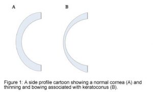
The cornea is the clear window of the eye, which allows light to be focused ontotheback of the eye. Keratoconus is a diseases that can compromise the integrity of the corneaby becoming misshapen, a process called ectasia. This predisposes to thinning and bulging of the cornea. Instead of having a curved ‘football’ shape it become pointed like a ‘rugby’ ball (Figure 1). This in turn increases astigmatism (when light is focused in different direction on to the back of the eye, the retina) and myopia (when an image is focused in front of the retina).
Who gets Keratoconus?
Both men and women may develop the condition, and usually this starts when we are teenagers or in our early twenties. The condition is problematic as it may continue to progress until vision becomes increasingly blurry, which may not be managed with spectacles or conventional soft contact lenses. Although inflammation of the surface of the eye or certain genetic conditions can result in keratoconus, usually a direct cause cannot be identified.
How do we diagnose Keratoconus?
Usually an optician will become suspicious that you have Keratoconus when they test your spectacle prescription. They may notice that you have become more myopic (short-sighted) or that your astigmatism has got worse. They may also notice that you have changes in your cornea that suggest you have Keratoconus. Normally they will refer you to see the ophthalmologist (eye doctor) who will also examine you and undertake special tests such as
- Topography (a painless test that measures how steep and thick your cornea is)
- OCT (a painless tests which measures the profile of the cornea)
- Specular microscope (a painless test that measures the health of the cells lining the back of your cornea, called the endothelium)
Sometimes the diagnosis is not clear and you may need to be monitored for a period to see whether the changes in your cornea become more apparent. We usually need to monitor you condition to decide if the disease is progressing (getting worse) and would benefit from treatment.
How can this be treated?
Keratoconus cannot be treated with eye drops or tablets. Often, and as long as we/your optician thin it is safe, we will try and improve your vision with contact lenses if glasses are of limited help to you. Contact lenses usually need to be hard (rigid), or occasionally scleral lenses to fit the shape of the cornea caused by keratoconus. Patients usually find contact lenses better than glasses as they allow light to enter the eye more normally, improving the quality of the image. If there is evidence that your disease is getting worse and it is safe we will consider arranging a treatment called corneal collagen cross-linking (CXL) to prevent the eye from warping further. Sometimes the cornea can become so thin that a break within causes fluid to build up which can be painful and suddenly causes clouding of vision. This is called hydrops and will need attention from the doctor quickly, involve specialist eye drops and can lead to scarring of the cornea.
What does corneal collagen cross-linking involve?
This is a treatment that aims to stiffen or harden the cornea and prevent it from becoming more misshapen. Although very long-term data is not available, this treatment has a good safety profile and low risk of complications such as infection. Further details can be discussed should you need this treatment but briefly it involves a day case procedure that requires the application of a specialist drops followed by ultra-violet light shone on the eye for several minutes.
Why might I need a corneal transplant?
Keratoconus can result in the cornea becoming so thin that CXL is not safe or likely to be effective, too pointed or scarred that KC cannot be managed by specialist contact lenses, you develop a perforation or when one cannot wear a contact lens. In these instances it may be necessary to replace part or all of your cornea through surgery by undertaking a corneal transplant called a graft or keratoplasty. This graft will come from a donor who has died and given consent for their cornea to be used for transplantation after their death.
There are three layers in the cornea:
- The epithelium (the front surface like a skin)
- The stroma (the middle part which makes up most of the cornea)
- The endothelium (the back surface which acts as a pump to stop the cornea from becoming waterlogged)
What types of corneal transplant are there?
Different diseases may affect all or part of the cornea. There are two broad approaches in keratoconus:
- Penetrating Keratoplasty (PK – a full thickness corneal transplant)
- Deep Anterior Lamellar Keratoplasty (DALK – a partial thickness corneal transplant replacing the epithelium and front part of the stroma up to 90% depth)
All involve the replacement of some or all of your cornea. Like all organ transplants, this involves taking corneal tissue from someone who has died and donated their organs for transplantation. Further information can be found in our leaflet concerning corneal transplantation.
Follow up and aftercare
Your surgeon will determine whether you have Keratoconus, whether it needs treatment with contact lenses, cross-linking or surgery. If you do not need treatment other than lenses we will usually monitor the condition every few months to see if the disease is progressing and you would benefit from further treatment.
Other advice:
- Stop wearing your usual contact lenses prior to and after your clinic visits as advised so that we can get an accurate scan.
- Contact the department as soon as you notice any problems e.g. increased blurring of vision, redness or pain that is getting worse. See an ophthalmologist as soon as possible if you start noticing these symptoms or any problems that may be of concern, so that the right treatment can be started straight away.
What is corneal cross-linking?
Corneal cross-linking is a minimally invasive treatment that strengthens the cornea, which is the clear, dome-shaped surface at the front of the eye.
It is most commonly used to treat progressive conditions called ectasia like keratoconus, where the cornea becomes weak and begins to bulge. The goal of CXL is to stabilise the cornea and prevent further thinning or distortion by reinforcing its internal structure.
How does corneal cross-linking work?
Corneal cross-linking works by strengthening collagen fibres in the cornea. The procedure involves applying riboflavin (vitamin B2) drops to the corneal surface, followed by exposure to ultraviolet (UV) light. This combination causes the collagen fibres to form stronger bonds, which stabilises the cornea and halts further thinning or bulging. The result is a more resilient cornea that is less prone to damage over time. The procedure is usually undertaken under local anaesthetic drops and typically takes around 30 minutes. Most patients generally return home the same day.
Benefits of corneal cross-linking
This treatment offers several important advantages, particularly for patients in the early stages of corneal disease. We will undertake a thorough assessment to determine whether there is evidence of the condition progressing and whether it is safe before recommending the treatment.
Prevention of disease progression
Keratoconus usually continues to progresses through to the age of 30-35, and the condition usually slows down by this age. The primary benefit of CXL therefore is its ability to slow down or stop the progression of corneal diseases in the early stages of keratoconus. By strengthening the cornea, the procedure prevents further thinning and deterioration, which in many cases prevents the need for a corneal transplant.
Improved visual stability
While CXL is not intended to directly correct refractive errors, it can stabilise the cornea, reducing fluctuations in vision. This stabilisation may lead to better visual clarity, especially in patients with early to moderate keratoconus. The condition is designed to be used in conjunction with other means of improving vision such as the use of specialist contact lenses.
Minimally invasive with quick recovery
The procedure is performed as a day case treatment (no overnight hospital stay required) with a relatively short recovery time. Most patients can return to their normal activities within a few days, although full visual improvement may take several months.
Long-term efficacy of corneal cross-linking
Corneal cross-linking has proven highly effective at slowing or halting the progression of keratoconus and similar conditions. Clinical studies have shown that the procedure can prevent further corneal damage for at least 5 to 10 years or more, and in many cases, only one treatment is needed.
However, CXL does not reverse existing damage to the cornea or restore lost vision. As a result, patients with advanced keratoconus, very thin corneas or significant scarring may still need additional treatment like corrective lenses or surgical procedures.
Side effects are usually mild and temporary. These may include light sensitivity, blurred vision, or discomfort in the first few days after treatment. Most patients recover without complications. Mr Williams will counsel you about the benefits and risks of the procedure and broadly these include discomfort in the first few days (for which we will prescribe analgesia), with less than 10% requiring retreatment and less than 3% having loss of vision due to rare complications such as poor healing, infection or scarring.
Corneal cross-linking provides an effective and minimally invasive way to try and halt the progression of corneal damage and preserve vision.
Pterygium
A pterygium is a growth of the conjunctiva (the membrane covering the white of the eye) on to the surface of the cornea (the window of the eye).
This often noticed when a patch may appear in he corner of one or both eyes. A pterygium is more commonly seen in hot climates, where exposure to sunlight is thought to play a role in its development. This is not always the case however and while occupations and recreations such as sailing and surfing may increase the risk, genetic factors play a role.
It is difficult to predict exactly who will develop a pterygium/pterygia (more than one pterygium). It usually develops slowly, over months or years. It may also cause episodes of redness or irritation. With time it can alter the profile of the cornea, leading to astigmatism (as the curvature of the eye becoming more flat) or cross the pupil. These can lead to a reduction in vision.
The treatment for pterygium is surgery. The aim is to remove the growth and create a barrier to prevent the pterygium from growing back. We do this by taking a healthy area of conjunctiva (usually from the top of the eyeball) in order to create a graft and move it to fill in the gap created. This is usually held in place by a harmless glue which dissolves after a few days or occasionally through stiches. This is enough time to allow the graft to heal. The eye will be red for a period of some weeks and will require drops to help reduce this inflammation and prevent infection.
All surgery carries risk including bleeding and infection. The risks of pterygium surgery include damage to one of the muscles surrounding the eye which is rare but can lead to double vision. The graft may also move following surgery and require further surgery. Very rarely an area of thinning may occur after surgery requiring additional treatment including surgery. There is a risk of high pressure during the period following surgery called glaucoma (high pressure in the eye which can usually be managed with drops).
Less than 10% of pterygia will return and this may require further surgery.
Blepharitis and Meibomian Gland Dysfunction (MGD)
Background
Normal tears of the eye are made up of three layers – an oily (lipid) layer, a watery (aqueous) layer and a sticky (mucous) layer.
There are Meibomian glands inside the eyelids with openings onto the edges of the eyelids (lid margins) which naturally produce oil. This oil stops the watery element of the tear film from drying out. Sometimes the glands become blocked (MGD) leading to tear film breakdown and ‘evaporative’ dry eyes. The glands may also produce excess oil which clogs the lashes causing crusting. You may experience discomfort, visual problems and other complications listed below.
Blepharitis is an inflammation of the eyelids causing irritation and discomfort. It can be persistent and usually affects both eyes.
It is common to have a combination of MGD, dry eye and blepharitis.
Symptoms of MGD and Blepharitis
- Sore eyelids – both eyes usually affected
- The eyes may feel gritty, itchy or as though they are burning
- The eyes may look inflamed or greasy
- The eyes may be sticky with discharge and stuck together in the mornings
- The eyes may be watery
- Flakes or scales may appear on the eyelids and eyelashes
- Glands may block and fill with oily fluid (chalazion)
Symptoms may come and go. It is common to have flare ups or long periods with no symptoms.
Possible Complications of Blepharitis
- Chalazion – swelling under the eyelid due to a meibomian cyst
- Stye – painful swelling outside the eyelid due to an infected eyelash
- Contact lens irritation
- Changes to eyelashes – loss of lashes, in growing lashes (trichiasis) or loss of colour to the eyelashes
- Eyelid ulceration and scarring
- Entropion (lid turning in) or ectropion (lid turning out)
- Conjunctivitis – sore red eye with discharge and/or watering
- Corneal ulceration and scarring
Treatment for MGD and Blepharitis
The aim of treatment is to control and manage the symptoms. There is no one off cure as inflammation tends to recur without treatment. Regular treatment will keep symptoms to a minimum. This treatment is mainly done by you.
Lid Hygiene
This is the most important part of your treatment.
The aim is to sooth the eyelids, unblock the meibomian glands and remove any stagnant oily secretions. This is achieved by cleaning the eyelids.
Warmth is used to break down the oil. The most successful method is a reusable bag device which can be heated in the microwave. It is preferable to a hot flannel as it retains heat longer (A hot flannel usually cools quickly and has to be constantly re-warmed in hot water to be effective). It should be applied for 5 -10 minutes.
Massage should be done immediately after applying the warmth. It helps to push the oil out of the glands. Massage along the length of the eyelids towards the eye (massage the upper lid down and the lower lid up) with the eyes closed using comfortable pressure. Massage for 30 seconds and repeat 5 – 10 times after warming.
Clean the eyelids following the warming and massage using a foam scrub as recommended by the doctor/nurse.
Repeat lid hygiene twice daily during a flare up. When symptoms subside continue as part of your daily routine.
If your symptoms are severe you may also be prescribed
- Antibiotics
- Artificial tears/lubricating drops/gel
Other advice
- Consider Omega 3 supplements – research shows that these may help relieve symptoms
- Avoid wearing eye make-up (especially eyeliner) when you have a flare up
- Avoid rubbing the eyes as it will make inflammation worse
- Continue your treatments for any other related conditions such as seborrheic dermatitis, rosacea or dry eye syndrome
- It is likely that unless you have severe dry eye complications or need for specialist drops your ongoing management will be with your optician or GP
About 1 in 10 patients may develop a film behind the lens implant following cataract surgery, often several years later. This is called posterior capsular opacification. It is usually amenable to laser treatment called ‘YAG laser capsulotomy’. This is undertaken on a microscope in clinic and creates an aperture behind the lens implant.
As with any intervention there are infrequently encountered risks. These include inflammation within the eye, high pressure, destabilising the lens (which rarely requires additional surgery), macula swelling add retinal damage. Should we recommend laser treatment, we will discuss these risks and prescribe drops in the first few days and weeks to reduce the chance of inflammation, high pressure and retinal damage.
Surgical Articles and Videos
All eye surgical procedures carry a level of risk including not obtaining the desired outcome through to varying levels of visual loss. Your eye surgeon will discuss the risks, benefits and alternatives of sight correction surgery, including those specific to your own circumstances, at the time of your preoperative consultation.
NHS Patients
- 01432 355444 ext 5898
- The County Hospital Hereford
- 2026 Geraint Williams
Private Patients
inc Hereford Vision Surgical Group
- Spire - 01905 350003
- Spire South Bank Hospital
- The Wye Clinic - 01432 802600